Authorised Indian Distributor
Saxsons Group
New Delhi, India · Since 1997
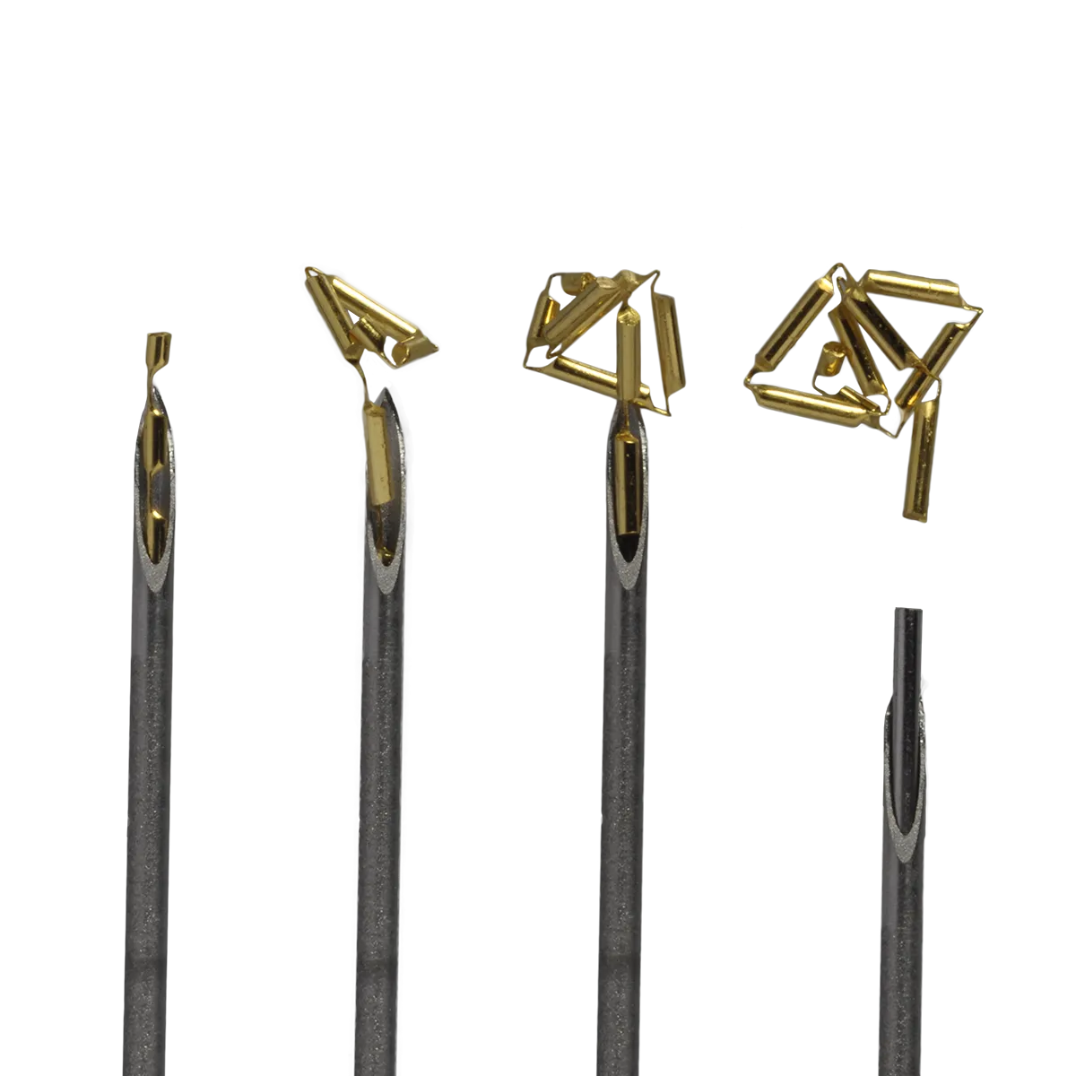
The Gold Anchor is a patented flexible gold-iron fiducial wire pre-loaded in an industry-leading thin needle. Multiple machined cut-outs cause the wire to fold on deployment — anchoring instantly in soft tissue for zero migration throughout the radiotherapy course. Visible on kV, MV, CBCT, CT and MRI, it enables precise image-guided and real-time tracking across the full spectrum of modern linac, particle and robotic radiotherapy platforms.
Patented cut-outs cause the marker to fold and mechanically lock in soft tissue the instant it exits the needle — confirmed stable across the full RT course.
Gold-iron alloy delivers strong contrast on kV, MV, CBCT, CT and MRI — the only flexible fiducial visible across every imaging modality used in modern radiotherapy.
Pre-loaded in an industry-leading thin needle (including 22G endoscopic) for EUS-guided pancreatic and bronchoscopic lung placements — minimal trauma, no anaesthesia typically required.
Compatible with SyncTraX, Varian Synchrony, CyberKnife and Elekta intrafraction management — enables continuous tumour tracking during beam delivery for liver, lung and prostate SBRT.
Clinically validated in proton and carbon-ion IGRT programmes. The gold-iron alloy provides high contrast imaging with negligible beam perturbation at clinical particle therapy energies.
Fully MRI-safe with no susceptibility artefact distortion at 1.5T and 3T. Enables MR-only treatment planning without CT co-registration — reducing workflow steps and imaging dose.
| Material | Gold-iron alloy wire (flexible, with machined cut-outs) |
| Design | Multiple cut-outs — folds and anchors on deployment |
| Imaging visibility | kV, MV, CBCT, CT, MRI (all modalities) |
| MRI status | MRI-compatible and MRI-safe |
| Delivery system | Pre-loaded in thin implantation needle (standard and endoscopic 22G) |
| Migration | Zero migration — patented anchor mechanism |
| Deployment locations | Prostate, liver, lung, pancreas, cervix, endometrium, esophagus, rectum, breast |
| Contraindications | Not for heart, large blood vessels, brain, spinal cord or eyes |
| Procedure time | 5–15 minutes (prostate); image guidance recommended |
| Permanence | Permanent (removable only by surgery) |
| Airport security | Not detected in routine security screening |
| Clinical evidence | 45+ peer-reviewed publications; prostate, liver, lung, pancreas, gynecologic |
| AERB / import status | Import licence required; Saxsons Group manages regulatory documentation |
Varian
TrueBeam Auto Beam Hold · On-board kV imaging
Elekta
XVI Seed Match · Intrafraction Motion Management
Accuray
CyberKnife Synchrony · TomoTherapy & Radixact MVCT
Brainlab
ExacTrac Dynamic® fiducial tracking
Hitachi / IBA / Varian
Proton therapy with real-time gating
HIMAC / NIRS
Carbon-ion radiotherapy with fiducial localisation
SyncTraX
Real-time tumour-tracking IGRT for liver SBRT
All platforms
CT/MRI fusion · MR-only planning workflows
Gold Anchor has been validated on Varian, Elekta, Accuray and Brainlab linacs, as well as proton and carbon-ion systems. It supports both CT-guided and MR-only planning workflows through its dual kV and MRI visibility.
Eight organ sites — one fiducial solution
The most widely published application. Gold Anchor markers are implanted transperineally or transrectally for daily kV or CBCT-guided prostate radiotherapy. Thin needles and instant anchoring make the procedure fast and well-tolerated.
Real-time tumour tracking in the liver using SyncTraX and Gold Anchor fiducials. The flexible anchor design maintains marker position despite respiratory motion — critical for liver SBRT dosimetric accuracy.
Transbronchial, CT-guided or endoscopic placement for peripheral and central lung lesions. Gold Anchor's thin needle minimises pneumothorax risk. Compatible with respiratory gating and real-time tracking.
Endoscopic ultrasound-guided (EUS) placement via 22G needle directly into the pancreatic tumour. Gold Anchor is a preferred choice for EUS-guided fiducial marking because its flexibility accommodates the endoscope trajectory.
Fiducial marking of cervical, endometrial and other gynaecological tumours for IGRT. MRI visibility enables MR-guided adaptive radiotherapy workflows without the need for secondary CT co-registration.
Post-biopsy clip marking to preserve lumpectomy site geometry. Also used as a radiation boost target marker in whole-breast IGRT, particularly for accelerated partial breast irradiation (APBI).
Pre-surgical marking of lung nodules, pancreatic tumours and other lesions to guide video-assisted thoracoscopic surgery (VATS) and laparoscopic resection. The visible marker eliminates intraoperative localisation uncertainty.
Used in proton and carbon-ion facilities for daily image-guided alignment. The gold-iron alloy provides high contrast on both kV imaging and MRI, with negligible particle therapy beam perturbation at clinical energies.
Conventional coil or cylindrical fiducials migrate because they rely on friction alone. Gold Anchor's patented cut-outs cause it to fold and mechanically lock in soft tissue the moment it exits the needle — confirmed stable in prostate, liver, lung and pancreatic SBRT series with follow-up imaging across the full treatment course.
Gold-iron alloy gives strong contrast on kV, MV, CBCT and CT while remaining MRI-compatible — rare for metallic implants. Departments running MR-only planning workflows can use the same fiducial without switching products. CT/MRI fusion-based planning is fully supported.
More peer-reviewed publications than any comparable flexible fiducial. Evidence spans prostate SBRT, liver tracking with SyncTraX, lung IGRT, EUS-guided pancreatic marking, gynecologic IGRT and particle therapy. Developed by the co-inventors of SBRT at Karolinska University Hospital — the clinical evidence base is built into the product's origin.
"The Gold Anchor has the thinnest needle and the best MRI visibility of any flexible fiducial I have used. It simply does not move once placed — which is exactly what you need for daily IGRT."
Radiation Oncologist, Tertiary Cancer Centre, India (Saxsons reference account)
Catalogs and brochures across the Gold Anchor range. White papers and clinical evidence live on the Knowledge Hub — link above.
Decide which Gold Anchor variant suits your clinical indication — diameter, length, alloy and needle gauge.
Full clinical brochure for prostate IGRT and SBRT — placement, imaging, dosimetry.
Multi-site fiducial marking — liver, lung, pancreas, gynecologic and more.
Post-biopsy clip and tumour-bed boost marker for whole-breast IGRT and APBI.
Implantation technique and customer testimonials from the clinical teams that use Gold Anchor every day. Educational and background videos are on the Knowledge Hub.
Gold Anchor™
Each card opens a focused post for a specific specialty — peer-reviewed evidence and clinical workflow, written for the persona you select.
For Radiation Oncologist
Conventional coil and cylindrical fiducials migrate up to 19–47 %. Can a 0.32 % migration rate change how prostate SBRT is planned?
Read this Radiation OncologistWhen the target moves with every breath, what does it take to get from 5.3 mm soft-tissue alignment error down to 1.5 mm?
Read this Radiation OncologistRoughly one in five standard coil markers migrate in lung parenchyma. What changes with a fold-anchor design and a thinner needle?
Read this Radiation OncologistWhat is the proton-fluence perturbation at clinical energies — and how do Gold Anchor fiducials compare to gold cylinders and titanium markers?
Read this Radiation OncologistA single-page overview of where the published evidence sits for prostate, liver, lung and particle therapy.
Read thisSaxsons Group supplies Gold Anchor fiducial markers across India with full import documentation, clinical in-service training and ongoing application support. Contact us to start a trial order or arrange a demonstration implantation workshop.